
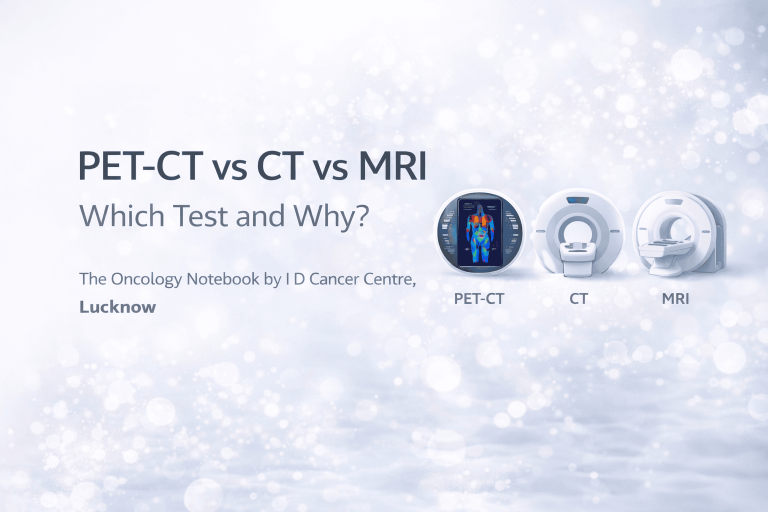
PET-CT vs CT vs MRI: Which Test and Why these Points Important In cancer Treatment?
When your doctor advises a scan—CT, MRI, or PET-CT—it is not “one scan is better than the other.” Each test answers a different clinical question. Choosing the right imaging at the right time improves diagnosis, staging, treatment planning, and follow-up—while avoiding unnecessary cost, delay, or radiation exposure.
This guide explains what each scan does, when it is preferred, and how to prepare.
1) Quick comparison (simple)
CT (Computed Tomography)
MRI (Magnetic Resonance Imaging)
PET-CT (Positron Emission Tomography + CT)
2) What each test actually “sees”
CT: detailed “X-ray slices”
CT uses X-rays to create cross-section images. It shows:
Organ size and shape
Lymph nodes (by size)
Lung nodules very well
- Bone structure wellOften done with contrast to highlight blood vessels and organs.
MRI: soft tissue and marrow clarity
MRI uses a magnetic field (no X-rays). It shows:
Brain and spinal cord details
Soft tissue planes (tumor vs muscle vs fat)
Marrow involvement
- Nerves, cartilage, pelvic organsOften enhanced with gadolinium contrast (different from CT contrast).
PET-CT: activity + location
PET uses a sugar-like tracer (commonly FDG) that active cells take up. Cancer often takes up more tracer. PET-CT shows:
Areas of increased metabolic activity
Whole-body spread (metastases)
- Helps differentiate scar vs active disease in selected settingsBut PET is not perfect—some infections/inflammation can also “light up.”
3) When CT is usually the first choice
CT is commonly used when doctors need a quick, comprehensive map of anatomy, such as:
Initial staging for many cancers (chest/abdomen/pelvis)
Lung evaluation (best general scan for lungs)
Suspected bowel obstruction, perforation, acute abdominal issues
Baseline imaging before systemic therapy
Assessing complications (pleural effusion, ascites, etc.)
CT is fast (minutes), widely available, and excellent for many initial assessments.
4) When MRI is the best choice
MRI is preferred when soft-tissue accuracy changes management, including:
Brain tumors or suspected brain metastasis
Spine (cord compression, nerve involvement)
Head & neck cancers (tumor extent in tongue base, nasopharynx, skull base, perineural spread)
Pelvic cancers (prostate, cervix, rectum) for local staging
Liver lesion characterization (in many cases)
Musculoskeletal tumors and marrow involvement
MRI often provides the most accurate local extent—useful for surgery planning and radiotherapy planning in selected cases.
5) When PET-CT is most helpful in cancer care
PET-CT is commonly used for:
Whole-body staging in many cancers when spread is suspected
Lymphoma staging and response assessment (very common indication)
Clarifying uncertain findings on CT/MRI (is a node/lesion active?)
Detecting recurrence when symptoms/tumor markers rise and CT is unclear
Post-treatment assessment in selected cancers (distinguishing scar from active disease), with correct timing
Important: PET-CT is not required for every cancer and every patient. It is most valuable when the result will change treatment decisions.
6) Head & neck cancer: practical examples
Because you treat many head & neck patients, this section is patient-friendly and clinically aligned.
CT is helpful for:
Neck nodes overview
Chest evaluation for lung spread (often CT chest)
Bone involvement in certain settings
MRI is helpful for:
Soft tissue extent (tongue base, nasopharynx, skull base)
Perineural spread and deep tissue planes
Better detail when CT is limited by dental artifacts
PET-CT is helpful for:
Whole-body staging in selected patients
Unknown primary (metastatic neck node with hidden primary) in some workflows
Post-treatment evaluation (timed appropriately) when residual disease is suspected
7) “Which is better?” is the wrong question
Examples:
“Is it cancer and where is it located?” → often CT/MRI + biopsy
“How far has it spread?” → CT or PET-CT depending on cancer type and scenario
“Is this lesion truly active disease or scar?” → sometimes PET-CT
“Is there brain involvement?” → MRI brain, not PET-CT
8) Preparation: what patients should know
CT preparation
If contrast CT: you may need kidney function test (creatinine)
Inform doctor if you have:
Prior contrast allergy
Kidney disease
Uncontrolled thyroid disease
Usually no fasting unless instructed.
MRI preparation
Inform staff if you have:
Pacemaker/implants (some are MRI safe, some are not)
Metal fragments (especially in eye)
Severe claustrophobia
Some MRIs need contrast; kidney function may be checked in specific patients.
PET-CT preparation (most important)
Typically fasting 4–6 hours (water allowed)
Avoid heavy exercise 24 hours before (can affect uptake)
Diabetes patients need special scheduling instructions
The scan requires resting quietly after tracer injection
Total visit time can be a few hours
9) Safety: radiation, contrast, and special situations
Radiation
CT and PET-CT involve radiation
MRI has no radiation
Contrast safety
CT contrast (iodine-based) can affect kidneys in vulnerable patients and can trigger allergic reactions in some.
MRI contrast (gadolinium-based) is different; risk considerations differ, especially in severe kidney disease.
Pregnancy and breastfeeding
Always inform your doctor and imaging center. Imaging choices may change.
10) Common reasons scans look “abnormal” but are not cancer
Patients often panic when a report shows something. Not all findings mean cancer:
Inflammation/infection can mimic cancer on PET-CT
Old healed infections can leave lung nodules/scars on CT
Benign cysts can appear in liver/kidney
Degenerative spine changes can look alarming but are common
This is why scans must be interpreted in clinical context by your treating team.
11) FAQs (patient-friendly)
Is PET-CT always the best for cancer?
No. PET-CT is very useful in specific scenarios, but many cancers are managed perfectly with CT and MRI plus biopsy.
Can PET-CT replace biopsy?
No. Imaging suggests cancer; biopsy confirms cancer and provides the type/grade/biomarkers.
Why did my doctor order CT first and later PET-CT?
Often CT gives the anatomical map first; PET-CT is added only if it will change staging or treatment.
Why MRI if I already had CT?
MRI may show soft tissue extent more accurately (brain, spine, pelvis, head & neck).
Can PET-CT show false positives?
Yes—some infections/inflammation show increased uptake. Your doctor correlates with symptoms, labs, and other imaging.
12) A practical “decision guide” (what doctors commonly do)
Initial evaluation / staging (many cancers): CT chest/abdomen/pelvis
Brain symptoms or high-risk cancers: MRI brain
Pelvic organ cancers (prostate/cervix/rectum): MRI pelvis for local staging
Lymphoma: PET-CT often for staging and response
Unclear lesion on CT/MRI: PET-CT can help assess activity
Post-treatment: imaging chosen based on cancer type, timing, and clinical need
Next steps at I D Cancer Centre
If you are confused about which scan is needed, bring:
All previous reports (CT/MRI/PET-CT)
CDs or digital images (not just the paper report)
Biopsy report and treatment summary (if already treated)
Our team will explain:
What the scan is meant to answer
Whether you need CT, MRI, PET-CT—or a combination
How results will influence your treatment plan
