
Leukoplakia: An Expert Guide to Causes, Cancer Risk, Diagnosis & Treatment
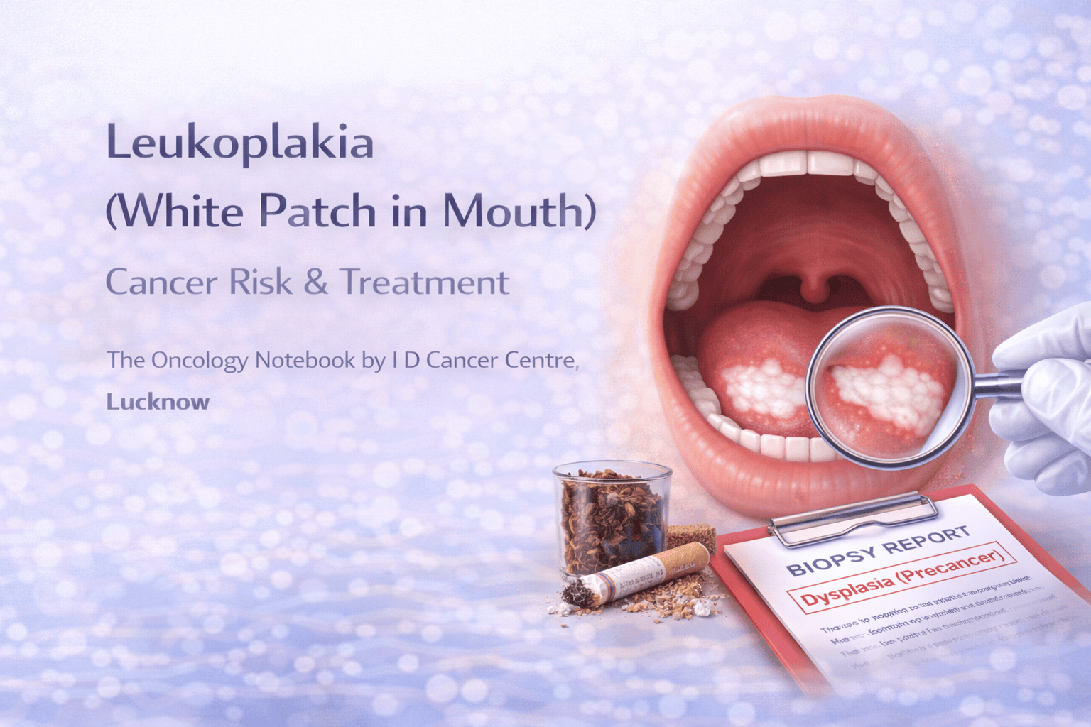
Leukoplakia is a common condition where a white patch appears on the lining of the mouth (and sometimes throat or voice box). Most leukoplakia patches are benign, but some can show pre-cancer changes (dysplasia) and may progress to oral cancer if ignored—especially in people who use tobacco or arealcohol.
The most important rule is simple:
Any white patch in the mouth that does not disappear in 2–3 weeks should be checked by a doctor/dentist and may need a biopsy.
This guide explains what leukoplakia is, how to recognize it, when it is dangerous, and what to do next.
1) What is leukoplakia?
Leukoplakia means a white patch or plaque on the mucous membrane that:
Cannot be rubbed off easily, and
Cannot be explained as another obvious condition (like thrush) at first glance
It is a clinical term, not a final diagnosis. Leukoplakia may represent:
Simple thickening of the lining due to irritation
Fungal or inflammatory changes
Pre-cancerous dysplasia
Early cancer (rare, but possible)
2) Where does leukoplakia occur?
Most commonly in the mouth:
Inside cheeks (buccal mucosa)
Tongue (especially sides)
Floor of mouth (under tongue)
Gums and inner lip
Soft palate
Less commonly, similar lesions can occur in:
Voice box (larynx)
Genitals (clinically different entities, but “white patch” concept exists)
3) How does leukoplakia look and feel?
Common appearance
White or grey patch
Thickened, rough, or wrinkled surface
May be flat or slightly raised
Often painless
Symptoms (if present)
Mild burning or irritation, especially with spicy food
Roughness felt by tongue
If ulceration or pain appears, risk becomes more concerning
Important: Oral thrush (fungal infection) usually rubs off leaving a red surface; leukoplakia typically does not.
4) What causes leukoplakia?
Leukoplakia is usually linked to chronic irritation and carcinogen exposure.
Major causes/risk factors (especially in India)
Tobacco chewing (gutkha, khaini, zarda, pan masala with tobacco)
Smoking (cigarette, bidi)
Alcohol (risk multiplies when combined with tobacco)
Areca nut (supari) (with or without tobacco)
Chronic friction: sharp tooth, ill-fitting denture, repeated cheek bite
Poor oral hygiene
Other contributors
HPV-related lesions are usually different (often papillomatous), but HPV is more relevant to oropharyngeal cancers; evaluation is clinical.
5) Is leukoplakia cancer?
Not always. Most leukoplakia is benign. But leukoplakia is important because it can be:
A pre-cancer lesion, or
A marker of “field change,” meaning the oral lining is exposed to carcinogens and at higher risk overall.
What increases the chance of pre-cancer/cancer?
Risk is higher when:
Patch is on the side of tongue or floor of mouth
Patch is non-homogeneous (mixed white-red, nodular, speckled)
There is ulceration, bleeding, or increasing pain
Patch is large or increasing in size
Person uses tobacco/areca nut or heavy alcohol
Age is higher and lesion is persistent
Previous oral cancer history
6) Types of leukoplakia (simple classification)
A) Homogeneous leukoplakia
Uniform white patch, flat, thin or mildly thick
Usually lower risk, but still needs evaluation if persistent
B) Non-homogeneous leukoplakia
Mixed pattern: white + red areas, nodular, verrucous, speckled
Higher risk for dysplasia and cancer
C) Erythroleukoplakia (red + white lesion)
Red components are more concerning and often warrant urgent biopsy.
7) When should you worry? (Red flags)
Seek evaluation promptly if you have:
White patch lasting > 2–3 weeks
Patch on tongue side or floor of mouth
Patch with red areas, nodules, or rough/irregular surface
Ulcer, bleeding, pain, or difficulty swallowing
Lump in neck, persistent sore throat, voice change (if lesion is deeper)
Tobacco/areca nut use history
8) How is leukoplakia diagnosed?
Step 1: Clinical oral examination
A clinician examines the mouth with good lighting and palpation.
Step 2: Rule out treatable causes
If thrush or obvious irritation is suspected:
Treat infection/irritation
Reassess in 2 weeks
Step 3: Biopsy (most important test)
If the patch persists or looks suspicious, a biopsy is recommended.
Biopsy tells:
No dysplasia (benign thickening)
Mild / moderate / severe dysplasia (pre-cancer)
Carcinoma in situ
Invasive cancer
Biopsy is the only way to confirm whether leukoplakia is pre-cancer or cancer.
9) Treatment: what actually works?
Treatment depends on biopsy and risk level.
A) Stop the cause (mandatory)
This is the most important step:
Stop tobacco, gutkha, pan masala, supari
Reduce/stop alcohol
- Fix sharp tooth/denture traumaMany lesions improve or stabilize after stopping the cause.
B) Regular follow-up (for low-risk lesions)
If biopsy shows no dysplasia or mild dysplasia and lesion is small:
Close observation with periodic oral exams
Photographic documentation may help
Repeat biopsy if lesion changes
C) Remove or ablate the lesion (selected cases)
If dysplasia is moderate/severe or lesion is high-risk:
Surgical excision
Laser ablation (in appropriate centers)
Other specialist-directed procedures
D) Treat associated conditions
Antifungals if candida is present
Manage nutritional deficiencies if identified
Improve oral hygiene
Important: “Mouth paints” or random long-term medicines should not replace biopsy-based management.
10) Can leukoplakia come back?
Yes—especially if tobacco/areca nut use continues. Recurrence is common without lifestyle change. Even after removal, new lesions can form because the oral lining remains exposed (“field cancerization”).
11) How can leukoplakia be prevented?
Avoid tobacco in any form (smoking/chewing)
Avoid areca nut/supari
Limit alcohol
Maintain oral hygiene
Fix dental irritants (sharp tooth, ill-fitting dentures)
Regular oral screening for high-risk individuals
12) Frequently asked questions
Is leukoplakia contagious?
No.
Can leukoplakia heal on its own?
Some lesions improve after removing irritants (especially stopping tobacco). But persistent lesions must be evaluated.
Is every leukoplakia pre-cancer?
No. But some are, and biopsy helps identify risk.
Does leukoplakia always turn into cancer?
No, but risk exists—especially in non-homogeneous lesions or when dysplasia is present.
Can I just use antifungal medicines?
Only if thrush is suspected and confirmed. If a patch persists, biopsy is needed.
Key takeaways (expert summary)
Leukoplakia is a persistent white patch that may be benign or pre-cancer.
Any patch lasting >2–3 weeks should be evaluated.
Biopsy is essential to assess dysplasia/cancer risk.
Stopping tobacco/areca nut is the most important treatment step.
High-risk lesions may need removal and close follow-up.
Evaluation at I D Cancer Centre
If you have a persistent mouth patch, oral ulcer, burning, or tobacco-related oral changes, early evaluation can prevent progression and detect oral cancer early.
